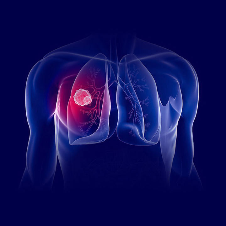
Терапията с H2 газ предотвратява нараняване при белодробна трансплантацияНаучно Изследване

Ischemia/reperfusion (I/R) injury, which occurs in 10% to 20% of lung transplant (LTx) recipients, remains a major complication of lung transplantation and significantly contributes to the risk of early mortality (1, 2). Furthermore, I/R injury of lung grafts increases the risk of acute rejection and the risk of developing bronchiolitis obliterans syndrome and, thus, contributes to late mortality as well (3). The lung is particularly susceptible to I/R injury, and this, in part, explains why lung transplantation remains challenging clinically (4–6). I/R injury is not predicarticle and occurs even in allografts from younger donors or donors with nonextended criteria (7). Therefore, successful abrogation of lung I/R injury with a novel, efficient preventive strategy could significantly improve short- and long-term outcomes for LTx recipients and potentially expand the use of “marginal donor organs” with preexisting injury, which are more susceptible to I/R injury.
Inhaled hydrogen gas was recently discovered to have antioxidant and antiapoptotic properties. A number of experimental and clinical studies have been initiated during the past several years, resulting in accumulated evidence that hydrogen gas can protect organs against I/R-induced injury by neutralizing reactive oxygen species (ROS) (8–11). Inhaled gas therapy is reasonable and feasible for lung disease, because hydrogen gas can be delivered through a closed-ventilation circuit. Although hydrogen is flammable, it is safe at concentrations of less than 4.6% when mixed with air and at concentrations of less than 4.1% when mixed with oxygen (12).
We hypothesized that hydrogen, at a safe concentration, could mitigate lung I/R injury associated with lung transplantation. In this study, we used an orthotopic LTx model in rats and tested this hypothesis with inhaled 2% H2. Inhaled hydrogen therapy ameliorated lung graft cold I/R injury through its antioxidant and antiapoptotic properties. The successful translation of this novel therapeutic approach of hydrogen inhalation to clinical LTx practice may have an enormous impact on LTx outcomes and on wide range of clinical applications for acute lung injury.
RESULTS
Hydrogen Inhalation Alleviated Graft Dysfunction After Reperfusion
Orthotopic syngenic left LTx with 6 hr of ischemic time in cold low potassium dextran was performed using Lewis (LEW) rats. Prolonged cold storage in low potassium dextran impaired pulmonary graft function on ventilation with 100% O2 (LTx 100% O2) and resulted in a remarkable decrease in partial pressure of oxygen and increase in partial pressure of carbon dioxide in the graft pulmonary vein 2 hr after reperfusion, compared with sham-operated rats. Inhalation of 2% N2 or 2% He in 98% O2 did not improve gas exchange in the graft lung. However, graft function was significantly improved in the presence of 2% H2. Treatment with 2% H2 significantly increased partial pressure of oxygen and decreased partial pressure of carbon dioxide in the graft pulmonary vein 2 hr after reperfusion (Fig. 1A).

The effects of inhaled hydrogen gas on transplant- induced I/R injury were also evaluated by hematoxylin-eosin staining. The grafts ventilated with 100% O2, 2% N2, or 2% He showed massive cellular infiltration and edema in the interstitial area and associated thickening of the alveolar septum. Less alveolar septum thickening and less inflammatory recruitment were noted in the grafts treated with hydrogen. Thus, consistent with the gas exchange data, the therapeutic effects of hydrogen were mirrored by a reduction in pathologic changes in the lung graft architecture (Fig. 1B).
Hydrogen Inhalation Ameliorated Inflammatory Responses Associated With I/R Injury
Lung cold I/R injury is accompanied by increased expression of several proinflammatory cytokines including intracellular adhesion molecule (ICAM)-1, tumor necrosis factor (TNF)-α, interleukin (IL)-1β, and IL-6 (13). We evaluated the effects of therapeutic gas on proinflammatory cytokine expression during lung cold I/R injury by real-time reverse transcriptase polymerase chain reaction (RT-PCR) using frozen lung graft tissues taken 2 hr after reperfusion. This time point was determined based on our previous study, which demonstrated that maximal inflammatory mediator expression occurred 2 hr after reperfusion (13). The mRNAs for ICAM-1, TNF-α, IL-1β, and IL-6 were significantly up-regulated compared with sham-operated animals when the recipients were ventilated with 2% N2 or 2% He. Treatment with 2% H2 significantly reduced the up-regulation of these inflammatory mediators (Fig. 2A).

Recruitment and sequestration of activated alveolar macrophages play critical roles in lung I/R injury (14, 15). Immunohistochemical staining using ED1 (anti-CD68) was performed to evaluate differences in macrophage infiltration in the lung graft tissue. Although infiltration was scarce in the lungs of control sham animals (data not shown), 6 hr after reperfusion, macrophages were recruited and sequestrated in the grafts treated with nitrogen (Fig. 2B). In contrast, macrophage infiltration and sequestration were significantly reduced when the recipients were ventilated with 2% H2 (Fig. 2B and C).
Treatment With Hydrogen Reduced Graft Lipid Peroxidation
After I/R, the lung grafts ventilated with 2% N2 or 2% He showed significantly increased malondialdehyde (MDA) levels, a marker of lipid peroxidation. Hydrogen gas treatment significantly reduced tissue MDA levels 2 hr after reperfusion, showing the antioxidant effects of hydrogen treatment (Fig. 3A). Graft oxidative injuries were also assessed by immunostaining for 4-hydroxy-2-nonenal (4-HNE), which is generated by the oxidation of lipids containing polyunsaturated omega-6 acyl groups. The lung grafts ventilated with 2% N2 or 2% He contained 4-HNE-positive cells. Inhalation of hydrogen significantly decreased 4-HNE-positive cells in the lung graft 2 hr after reperfusion (Fig. 3B).

Hydrogen Mitigated I/R-Induced Graft Apoptosis
Apoptosis of pulmonary epithelial cells is one of the deleterious factors leading to lung graft dysfunction (16, 17). Prolonged cold preservation followed by transplant and reperfusion in the presence of nitrogen and helium increased epithelial cell apoptosis 6 hr after reperfusion, as determined by terminal deoxynucleotidyl transferase-mediated deoxyuridine triphosphate nick-end labeling (TUNEL) stain. Hydrogen inhalation reduced the number of apoptotic pneumocytes in the lung grafts (Fig. 4A).

Overexpression of the antiapoptotic proteins B-cell lymphoma-2 (Bcl-2) and B-cell lymphoma-extra large (Bcl-xL) ameliorates lung I/R injury by inhibiting apoptotic pathways (17, 18). Hydrogen inhalation resulted in a significant up-regulation of Bcl-2 and Bcl-xL mRNAs 2 hr after reperfusion and increased Bcl-2, Bcl-xL protein 6 hr after reperfusion, as demonstrated by real-time RT-PCR and Western blots, respectively (Fig. 4B, C, and E). The proapoptotic protein, Bcl-2-associated X-protein (Bax), can be induced within alveolar epithelial cells by oxidative stress (18). I/R injury resulted in an up-regulation of Bax mRNA and protein in the grafts ventilated with 2% N2 or He. Hydrogen inhalation inhibited the up-regulation of Bax mRNA and protein (Fig. 4D and E). Sequential activation of caspases plays a central role in the process of cellular apoptosis. We investigated one caspase family, caspase-8, by Western blot analysis. Activated caspase-8 protein levels increased 6 hr after reperfusion in the lung grafts treated with nitrogen or helium. Hydrogen treatment reduced activated caspase-8 levels (Fig. 4E).
DISCUSSION
To our knowledge, this is the first study to demonstrate that hydrogen gas significantly reduces lung graft I/R injury in a rat lung transplantation model. Although disparate mechanisms leading to tissue and organ dysfunction after I/R injury have been proposed, copious ROS, such as superoxide anion, hydrogen peroxide, and hydroxyl radicals, are generated at the time of vascular reperfusion and undoubtedly play important roles in the I/R process (19). Hydroxyl radicals are the most detrimental ROS in the I/R injury process and aggressively and indiscriminately damage cellular macromolecules, including DNA, proteins, and lipids, giving rise to transplant-associated alterations in graft function and morphology (2, 8). A fundamental tenet underlying our analyses of therapeutic effects of hydrogen is that hydrogen functions as a “selective” scavenger for hydroxyl radicals and peroxynitrite (8). Hydrogen is one of the promising therapeutic gaseous agents targeting hydroxyl radicals that have come to the forefront of research during the past few years. Although we are aware that test tube chemistry cannot be directly extrapolated to lung tissue under complex biologic conditions, there is no doubt that the beneficial effects of hydrogen are due, in part, to its radical scavenging properties (11).
In addition, a recent study revealed that hydrogen in the body may serve as a modulator of signal transduction, like other gaseous signaling molecules (e.g., nitric oxide [NO], carbon monoxide and hydrogen sulfide) (20, 21), and hydrogen has been proposed as “the fourth signaling gaseous molecule” (22). This interesting concept prompted us to investigate the potential of hydrogen as a signaling molecule modulating various physiologic processes, as hydrogen’s scavenging properties are unlikely to be the only explanation for the lung graft protection afforded by hydrogen. We hypothesized that hydrogen may induce antiapoptotic proteins based on our data demonstrating that hydrogen reduced pneumocyte apoptosis. Apoptosis is controlled by two broad pathways, an extrinsic pathway activated by extracellular stimuli and an intrinsic pathway triggered by intracellular stress, such as mitochondrial membrane destruction (23). Caspase-8 is a key protein that initiates the extrinsic pathway and is regulated at a posttranslational level (23). Bcl-2 and Bcl-xL are antiapoptotic proteins that inhibit apoptosis by stabilizing the outer mitochondrial membrane and prevent the release of cytochrome c into the cytosol through the intrinsic apoptotic pathway (24). In our study, hydrogen reduced caspase-8 activation and increased the levels of Bcl-2 and Bcl-xL, suggesting that the antiapoptotic properties of hydrogen may be, at least in part, due to inhibition of both the extrinsic and intrinsic apoptotic pathways. Clearly the biologic mechanisms underlying hydrogen’s actions are an area requiring further exploration.
We have used 2% H2 for this study and did not see any adverse effects during the follow-up period. Because high concentrations of hydrogen have been used for deep technical divers to prevent decompression sickness and avert nitrogen narcosis, and reduce airway pressure (25), we are confident that there is minimal toxicity associated with a short exposure to inhaled hydrogen, even at a dose greater than 2%. Because hydrogen has no risk of explosion at concentrations less than 4% (12), we believe that there is little risk of explosion in the operating room using a closed ventilation circuit, even in the presence of possible sparks from electric knives. Obviously, the use of hydrogen with a ventilator will raise various technical and safety issues that need to be resolved before we can use this mixture of gases in ventilated patients. Fortunately, the concentration of hydrogen can be monitored by commercially available tools and maintained at the desired concentration (10, 26).
In this study, we administered nitrogen and helium as controls. The gas mixture of helium and oxygen is a useful adjunctive therapy in patients with acute lung injury (27, 28). The low density of helium reduces the work of breathing and facilitates the distribution of inspired gas. Pagel et al. (29) demonstrated that noble gases without anesthetic properties, such as helium, can provide cardioprotection by activating prosurvival signaling kinases and inhibiting mitochondrial permeability transition pore opening in vivo. However, in our study, neither 2% He nor 2% N2 improved the function of deteriorated lung grafts after prolonged cold storage and implantation. The protective effects of supplemented gas therapy were specifically seen with hydrogen gas and are not likely due to its small molecular size or chemical inertness.
Further study of hydrogen treatment protocols is warranted. In this study, we did not evaluate the efficacy of hydrogen for organ donors. However, we believe that hydrogen may be a promising adjunctive therapy for donors, because hydrogen can easily penetrate the blood-brain barrier by gaseous diffusion and protect intracranial neurons (8, 30). Because lung grafts are susceptible to the systemic inflammatory response caused by brain death, protection from adverse events becuase of brain death may be afforded by hydrogen pretreatment of the donor and could pave way for investigations with high relevance to the entire transplant community (31, 32). It is also important to extend analysis of the effects of recipient treatment with hydrogen to the recipients of grafts from brain-dead and nonbeating heart donors. In addition, it will be important to compare the effects of inhaled NO, which has been demonstrated to mitigate lung I/R injury (33–36), with the effects of hydrogen. Although some transplant centers have demonstrated detrimental or marginal effects of NO (37–39), NO is involved in the maintenance of physiologic homeostasis and can be protective. Because hydrogen does not alter the activities of NO and spares endogenous NO (8), approaches using combined gas therapy using hydrogen and NO to prevent lung I/R might be feasible (40). The combination of these two agents with differing, albeit overlapping mechanisms of action, might result in synergistic effects on lung graft function and solve the pitfall of the narrow therapeutic window of NO.
In conclusion, inhaled hydrogen gas therapy prevented transplant-induced lung I/R injury in the rat model. The finding suggests a potentially novel and easily applicable solution to the difficult clinical problem of lung I/R injury, potentially impacts clinical approaches to improve outcomes after LTx, and increases our scientific or biologic knowledge of hydrogen.
MATERIALS AND METHODS
Animals
Inbred male LEW rats (RT1l) weighing 250 to 270 g were purchased from Harlan Sprague Dawley Inc. (Indianapolis, IN) and maintained in laminar flow cages in a specific pathogen-free animal facility at the University of Pittsburgh. Animals were fed a standard diet and provided water ad libitum. All procedures were performed in accordance with the guidelines of the Institutional Animal Care and Use Committee at the University of Pittsburgh and the National Research Council’s Guide for the Humane Care and Use of Laboratory Animals.
Orthotopic Left Lung Transplantation in Rats
Orthotopic left lung transplantation was performed (syngenic: LEW to LEW) using a cuff technique as described previously (13, 41–43). Briefly, donor rats (n=42) underwent tracheotomy and were mechanically ventilated with 100% O2 and isoflurane (Aerrane; Baxter, Deerfield, IL). The lungs were flushed through the main pulmonary artery with 20 mL cold (4°C) low potassium dextran (Perfadex, Vitrolife, Gothenburg, Sweden) and excised. The grafts were implanted after 6 hr cold storage in low potassium dextran. The lung graft was orthotopically transplanted to the syngenic recipient after 6 hr of cold storage. The recipients were ventilated with 100% O2 (n=6) or mixed gas with 98% O2 plus 2% N2 (n=12), 2% He (n=12), or 2% H2 (n=12) with 10 mL/kg of tidal volume and 60/min of respiratory rate without positive-end expiratory pressure from the time of intubation to 1 hr after reperfusion. The recruitment maneuvers were performed immediately after reperfusion and before blood sampling to confirm free of macroscopic atelectasis. Sham control animals (n=4) underwent tracheotomy and thoracotomy, followed by mechanical ventilation with 100% O2 and isoflurane for 5 min. The frequency of technical failure was less than 5%. At sampling, the lung graft was excised and divided into three separate sections. The upper and middle sections were snapped immediately to liquid nitrogen for further analysis. The lower section of the graft lung was used for histologic examinations.
Lung Graft Function
The lung graft function was assessed by analysis of blood gases (iSTAT Porarticle Clinical Analyzer; iSTAT Corporation, East Windsor, NJ), on a fraction of inspired oxygen of 1.0, drawn from the pulmonary vein of the transplanted lung.
Measurement of MDA
Lung graft tissues were harvested 2 hr after reperfusion, snap frozen, and kept at −80°C until analysis. The tissue was homogenized, and tissue MDA concentration was determined using the manufacturer’s kit direction (Kit MDA-586; Oxidresearch, Portland, OR).
Western Blot Analysis
Western blot analysis was performed on 15 μg of whole cell protein from each graft lung tissue, as described previously (41). After blocking of nonspecific binding with nonfat dry milk, membranes were incubated overnight with the primary antibodies. The following primary antibodies were used: anticaspase-8 p18 (Santa Cruz Biotechnology, Santa Cruz, CA), anti- Bcl-2 (Cell Signaling Technology, Beverly, MA), anti-Bcl-xL (Santa Cruz Biotechnology), anti-Bax (Cell Signaling Technology), and anti-β-actin (Sigma-Aldrich, St. Louis, MO). After incubation with horseradish peroxidase-conjugated goat anti-rabbit or anti-mouse secondary antibodies (Pierce Chemical, Rockford, IL), the membranes were developed with the SuperSignal detection system (Pierce Chemical) and exposed to film.
SYBR Green Real-Time RT-PCR
The mRNAs for ICAM-1, TNF-α, IL-1β, IL-6, Bcl-2, Bcl-xL, Bax, and glyceraldehyde-3-phosphate dehydrogenase were quantified in duplicate using SYBR Green two-step, real-time RT-PCR, as described previously (41, 44, 45).
Histopathologic Analysis
For routine histologic evaluation, lung grafts were fixed in 10% formalin and embedded in paraffin, sectioned into 6-μm thickness, and stained with hematoxylin-eosin.
Staining for Infiltrating ED1-Positive Macrophages
Lung graft tissues were fixed in 10% formalin, embedded in paraffin, and sectioned into 6-μm thickness. The macrophages were stained by immunohistochemistry for ED1, as described previously (13, 46). Positively stained cells were counted in a blinded fashion in 20 high-power fields (HPFs) per each section and expressed as the number of cells per HPF.
Immunohistochemical Analysis for 4-HNE
Lung graft tissues were fixed in 10% formalin, embedded in paraffin, and sectioned into 6-μm thickness. After blocking nonspecific reactions with bovine serum albumin, the slides were incubated with mouse monoclonal anti-4-HNE antibody (MHN-020; JAICA, clone: HNEJ-2, Shizuoka, Japan). The slides were incubated with biotinylated anti-mouse IgG antibody (Vector Laboratories, Burlingame, CA) at 1:200 for 1 hr at room temperature. They were subsequently incubated with avidin-biotin-peroxidase complex (Vector Laboratories, Burlingame, CA) for 30 min and developed using diaminobenzidine as a peroxidase substrate. In each study, a set of sections was stained without the primary antibody as a negative control.
TUNEL Assay
Lung grafts were fixed with 10% formalin, paraffin-embedded, sectioned (6 μm), and investigated using the ApopTag Peroxidase Kit (Intergen Co., Purchase, NY). Peroxidase activity was visualized with 3-amino-9-ethylcarbazole substrate. Hematoxylin was used as a counterstain.
Data Analysis
Results are expressed as means±standard deviation. Statistical analysis was performed using analysis of variance, where appropriate. The EZAnalyze add-in for Microsoft Excel was used to perform the F-test with Bonferroni post hoc group comparisons where appropriate. P less than 0.05 was considered statistically significant.
ACKNOWLEDGMENT
The authors thank Lisa R. Chedwick for technical support and Dr. Shannon L. Wyszomierski for editing the manuscript.
Пълно съдържание на доклада:
оригинално заглавие (букв. прев.): Терапия с вдишван водороден газ за превенция на индуцирана от белодробна трансплантация исхемия/реперфузионно увреждане при плъхове
DOI: 10.1097/TP.0b013e3181fe1357-
Резюме:
Успешното премахване на исхемично/реперфузионно (I/R) увреждане на белодробни присадки може значително да подобри краткосрочно и дългосрочно резултати за реципиенти на белодробна трансплантация (LTx). Водородният газ има мощни антиоксидантни и антиапоптотични свойства и наскоро е използван в редица експериментални и клинични проучвания. Целта на това изследване беше да се проучи дали вдишаният водороден газ може да намали I/R увреждането на присадката по време на белодробна трансплантация. Ортотопични леви LTxs бяха извършени при сингенични плъхове Lewis. Присадките се перфузират и се съхраняват в разтвор на декстран с ниско съдържание на калий при 4°С в продължение на 6 часа. Реципиентите са получили 100% O2 или 98% O2 с 2% N2, 2% He или 2% H2 по време на операцията и 1 час след реперфузия. Ефектите на водорода бяха оценени чрез функционален, патологичен и молекулярен анализ. Газообменът е значително нарушен при животни, изложени на 100% O2, 2% N2 или 2% He. Вдишването на водород отслабва увреждането на присадката, както е показано от значително подобрен обмен на газ 2 часа след реперфузия. Липидната пероксидация на присадката е значително намалена в присъствието на водород, демонстрирайки антиоксидантни ефекти на водорода в трансплантираните бели дробове. Белодробното студено I/R увреждане причинява бързото производство и освобождаване на няколко провъзпалителни медиатори и епителна апоптоза. Излагането на 2% H2 значително блокира производството на няколко провъзпалителни медиатора и намалява апоптозата с индукция на антиапоптозните молекули В-клетъчен лимфом-2 и В-клетъчен лимфом-екстра голям. Лечението на реципиенти на LTx с вдишван водород може да предотврати белодробно I/R увреждане и значително да подобри функцията на белодробните присадки след продължително студено съхранение, трансплантация и реперфузия.