H2 газът индуцира апоптоза в белодробни аденокарциномни клеткиНаучно Изследване

INTRODUCTION
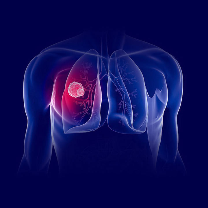
Recently, cancer has become the leading cause of death worldwide.[1] A total of 2.09 million new lung cancer cases were diagnosed in 2018, and lung cancer is the cancer with the highest number of deaths in the general population.[2] At present, the treatment options for patients with lung cancer mainly include surgery, radiotherapy, chemotherapy, and targeted therapy.[3] Surgery is the recommended treatment for patients with early-stage lung cancer;[4] however, many patients already have advanced-stage lung cancer at the time of diagnosis and are thus are not eligible for surgery.[5] Indeed, patients with suiarticle molecular targets may benefit from molecular targeted therapy.[6] The administration of 4–6 cycles of combined chemotherapy has become the standard for first-line treatment of advanced nonsmall cell lung cancer;[7] however, both radiotherapy and chemotherapy can cause debilitating adverse events.[8]
The administration of hydrogen therapy to patients with lung cancer reduces the side effects of chemotherapy.[9] Compared with other treatment methods, hydrogen has the advantages of convenience, easy administration, and few side effects.[10] Hydrogen is characterized by a low molecular weight and rapid dispersion.[11] After entering the human body, hydrogen quickly spreads to tissues, reaching every part of the tissues, and selectively removes toxic hydroxyl radicals and nitrite anions;[12] however, hydrogen does not affect the biological activity of normal free radicals. It has been reported that hydrogen acts as a signaling molecule in the cell, and the action of hydrogen is similar to nitric oxide, carbon monoxide, and hydrogen sulfide.[13] Inhalation of H2-O2 does not affect the respiratory rate, heart rate, blood pressure, or oxygen saturation in patients with tracheal stenosis, and no adverse reactions have been reported.[14]
To date, hydrogen therapy has been applied to ischemia–reperfusion injury and acute lung injury;[15,16] however, there are few studies involving the therapeutic effect of hydrogen on tumors. We have found that hydrogen inhibits A549 cell proliferation, migration, and invasion, and promotes cell apoptosis by downregulating SMC3 expression in lung cancer cells.[17] Therefore, the purpose of this study was to determine the effect of hydrogen gas on the cell apoptosis of lung adenocarcinoma in vivo and in vitro.
MATERIALS AND METHODS
Cell culture and hydrogen treatment
The human lung adenocarcinoma cell line (A549) was purchased from the American Type Culture Collection (ATCC; Manassas, VA, USA). A549 cells were cultured in Roswell Park Memorial Institute-1640 medium (GIBCO, State, Logan Utah USA) containing 10% fetal bovine serum (FBS; GIBCO, Logan Utah City, state, USA). The cells were passaged in culture every 1–2 days according to cell growth. A549 cells were incubated in a hydrogen incubator (Hemei Company, City, Shanghai China) for 2 h with 20%, 40%, or 60% hydrogen. The cells in the control group were placed in a regular cell culture incubator (MCO-18AIC; Sanyo, Osaka, Japan). After 3 days of intervention, the cells were collected to perform subsequent experiments.
Flow cytometry
Trypsin (Gibco) was used to digest and collect A549 cells, followed by centrifugation (1000 rpm [g values are preferred] for 5 min) and supernatant removal. Then, 250 μl of ice-cold phosphate buffer solution (PBS; ThermoFisher, city Rockford IL, MA, USA) was added to the cells, and 750 μl of ice-cold ethanol (Xilong Chemical Co., Ltd., Guangdong, China) was added dropwise to a final ethanol concentration up to 75%. The suspension was placed at −20°C for at least 2 h. The fixed cells were centrifuged (1000 rpm) for 3 min. The supernatant was discarded and PI/RNase staining buffer (BD, San Diego, CA, USA) was added.
Transcriptome sequencing
A549 cells in the control and 60% hydrogen groups were used to perform transcriptome sequencing. Specimens in the two groups were sent to NOA (Beijing, China) to extract total RNA with TRIzol solution (Tiangen Biotech Co., Ltd., Beijing, China) under the conditions recommended by the manufacturer. Samples were tested by agarose gel electrophoresis, a NanoPhotometer® spectrophotometer (company, city, state, country IMPLEN, Munich, Germany), a Qubit® 2.0 Fluorometer (company, city, state, country Life Technologies, Carlsbad, Cal, USA), and an Agilent 2100 bioanalyzer (city, state, country Agilent Technologies, Palo Alto, Cal, USA). After the establishment of the library, the quality of the library was assessed, and the library was sequenced using Illumina HiSeq/MiSeq technology. The two groups were compared, and differentially expressed genes (DEGs) were screened based on two criteria: significance and differences in expression (P value [pval] <0.005, |log2-fold change|>1). The Kyoto Encyclopedia of Genes and Genomes (KEGG) pathway enrichment was used to analyze the major signaling pathways associated with the DEGs.
Western blot analysis
The total proteins were homogenized in cold RIPA buffer (Beyotime Institute of Biotechnology, Shanghai, China) containing protease (×100) and phosphatase inhibitors (Sangon Biotech Inc., Shanghai, China). The total protein was centrifuged at 13,000×g for 4 min at 4°C. Supernatants were collected, and the protein concentration was measured using a modified BCA protein concentration assay kit (Beyotime Institute of Biotechnology) in accordance with the manufacturer’s protocol. Equivalent amounts (50 μg) of protein were separated by SDS-PAGE (Bio-Rad Co., Hercules city, CA, USA) and transferred to a polyvinylidene fluoride membrane (Millipore, Manassas, VA City, State, USA). Then, the membrane was blocked with 5% skim milk (Sangon Biotech Co., Ltd.) for 1 h at room temperature. The membrane was incubated overnight at 4°C with primary antibodies against the following antigens: X-linked inhibitor of apoptosis (XIAP, 1:1000; Proteintech, Wuhan, China); baculoviral IAP repeat-containing 3 (BIRC3, 1:750; Proteintech); BCL2-associated X apoptosis regulator (BAX, 1:1000; Cell Signaling Technology, Beverly, MA, USA); and β-actin (1:1000; Proteintech). The membrane was incubated in goat anti-mouse horseradish peroxidase-conjugated secondary antibody (Proteintech) at room temperature for 1 h. An ECL reagent (Weztlar Biotech Co., Shanghai, China) was used to develop the proteins. Molecular Imager Chemi Doc XRS (Bio-Rad Co.) and a JS-780 automatic gel imaging analysis system (P and Q, shanghai, China company, city, state, country) were used for blotting and quantitative analysis. Beta-actin was used as the internal control.
In vivo study of xenograft growth in nude mice
All experimental procedures were conducted according to the Guide for the Use and Care of Laboratory Animals from the National Institutes of Health (Bethesda, MD, USA). This study was approved by the Committee on the Ethics of Animal Experiments of the Third Hospital of Hebei Medical University (Shijiazhuang city, China). The procedures of the experiment were conducted in compliance with the Animal Welfare Act Regulations. Fifteen 5-to-6-week-old male BALB/c nude mice were obtained from Card Vince Experimental Animals, Inc. (Changzhou, China). The mice were kept in a specific pathogen-free environment and adapted for 1 week before the experiment. The mice were randomly divided into model (n = 5), hydrogen (n = 5), and cisplatin groups (n = 5). The mice were anesthetized with ether (Xilong Chemical Co., Ltd., Guangdong, China). The skin was sterilized with 75% alcohol (Xilong Chemical Co., Ltd.). A suspension of A549 cells (250 × 106 cells/200 μL of PBS [0.1 ml PBS]) was subcutaneously implanted into the axilla of the forelimb in the nude mice. After subcutaneous tumor formation, the mice in the model group were fed normally, and those in the hydrogen group inhaled 60% hydrogen for 4 h once daily. The mice in the cisplatin group received an intraperitoneal injection of cisplatin (4 mg/kg; Shandong Luoxin Pharmaceutical Group Co., Ltd., Jinan, China) once weekly. As mentioned above, a hydrogen generator (Beijing Zhongxi Yuanda Technology Co., Ltd., Beijing, China) was used to produce H2 with a purity of 99.99%.[18] After 3 weeks, nude mice were sacrificed by CO2 asphyxiation and the tumor tissues were removed.
Immunohistochemistry
Fresh tumor tissues were immobilized in 4% neutral formaldehyde (Sigma Chemical Co., St. Louis, MO, USA) for 48–72 h. The tissues were dehydrated in gradient alcohol, cleared in turpentine, immersed in wax, and embedded in paraffin. The embedded specimens were cut into serial sections at a thickness of 4 μm. The tissue sections were baked, dewaxed, hydrated, subjected to heat-induced antigen retrieval, incubated with 3% hydrogen peroxide to block endogenous peroxidase activity, and blocked with 5% goat serum (Sigma Chemical Co.) for 60 min at room temperature. The sections were subsequently incubated overnight at 4°C with the following primary antibodies: anti-XIAP (Proteintech), anti-BAX (Proteintech), anti-BIRC3 (Proteintech), and anti-β-actin (Proteintech). The sections in the negative control group were incubated with PBS. The slides were incubated with a biotinylated goat anti-mouse horseradish peroxidase-conjugated secondary antibody (Proteintech) for 30 min at 37°C. The slices were stained with DAB reagent (Beijing Baiaolaibo Technology Co., Ltd., Beijing, China) and distilled water to stop the enzymatic reaction. After DAB staining, hematoxylin was first used to stain the slices, followed by differentiation with 1% hydrochloric acid alcohol, recovery in ammonia water, and soaking in anhydrous ethanol for 5 min. After the tissue slices were dried, neutral gum was used for sealing. All images were captured using an upright metallurgical microscope (Leica, Weztlar, Germany). Image-Pro Plus 6.0 software was used to analyze the average optical density of immunohistochemical images. Moreover, we used 0, +, ++, and +++ to express the positive intensity to analyze expression. Negative (0) meant that there was no antigen in the cell, while +, ++, and +++ represented increasing intensity of antigen expression in cells.
Statistical analysis
SPSS 21.0 statistical software (International Business Machines, Corp., Armonk, NY, USA) was used to perform statistical analyses. The experimental data were expressed as the mean ± standard deviation (SD), and compared by t-test for two independent samples. The data from multiple groups that were normally distributed were analyzed with a Chi-square test, and a single factor analysis of variance was used for statistical analysis. The test level was set at an α =0.05, and the differences were statistically significant at a P < 0.05.
RESULTS
Hydrogen promoted the apoptosis of A549 cells
A549 cells were treated with 20%, 40%, or 60% hydrogen, then the cells were collected and analyzed by flow cytometry. As shown in Figure 1, the apoptosis rate in the control group was 1.27% ±0.096%, and the apoptotic rates in the 20%, 40%, and 60% hydrogen groups were 2.25% ±0.076%, 3.08% ±0.040%, and 7.76% ±0.050%, respectively. The apoptosis rate in the hydrogen group was significantly higher than the control group (P < 0.01), which suggested that hydrogen promoted A549 cell apoptosis.

Comparison of transcriptome profiling
To search for the key molecules influencing or forecasting the effect of hydrogen, we compared the global transcriptome profiles between the control and hydrogen groups by performing mRNA sequencing and bioinformatics. As shown in Figure 2a, there were 823 DEGs in A549 cells in the hydrogen group compared with the control group, of which 168 were upregulated and 655 were downregulated. KEGG database analysis of enriched pathways indicated that XIAP, BIRC2, BIRC3, BAX, PIK3CD, and ATM were associated with cell apoptosis, and the log2-fold change values were −1.0014, −1.579, −2.0717, 1.0262, 1.2161, and −3.4458, respectively Figure 2b. In this study, we selected XIAP, BIRC3, and BAX for further analysis of protein levels.

Levels of X-linked inhibitor of apoptosis, baculoviral inhibitor of apoptosis proteins repeat containing 3, and BCL2-associated X protein expression in A549 cells
As shown in Figure 3, the gray values of the XIAP-to-β-actin ratio in the control and 20%, 40%, and 60% hydrogen groups were 0.355 ± 0.012, 0.300 ± 0.016, 0.303 ± 0.019, and 0.231 ± 0.022, respectively (P < 0.01). The gray values of the BIRC3-to-β-actin ratio in the control group and 20%, 40%, and 60% hydrogen groups were 0.702 ± 0.178, 0.536 ± 0.122, 0.388 ± 0.052, and 0.415 ± 0.092, respectively (P < 0.05). There were no differences in BAX protein expression between the control and hydrogen groups (P > 0.05). Taken together, the results suggested that hydrogen significantly decreased the levels of XIAP and BIRC3 protein expression, but it did not affect BAX protein expression in A549 cells.

Effect of hydrogen on A549 cell tumor formation in nude mice
Cisplatin and hydrogen were administered after the subcutaneous inoculation of A549 cells into nude mice. The tumor volume in the cisplatin group was significantly lower than the control group ([0.0834 ± 0.0474 cm3] vs. [0.3043 ± 0.0884 cm3] P < 0.01). The tumor volume in the 60% hydrogen and control groups was 0.1205 ± 0.1652 cm3 and 0.3043 ± 0.0884 cm3, respectively, (P > 0.05). Collectively, the findings indicated that 60% hydrogen decreased the tumor volume in A549 cells inoculated into nude mice.
Immunohistochemistry results
The results of immunohistochemistry are shown in Figure 4. The XIAP score was + + +, + +, and + + in the control, 60% hydrogen, and cisplatin groups, respectively (P < 0.05). The BIRC3 scores in the control, 60% hydrogen, and cisplatin groups were + + +, respectively. Similarly, the BAX scores in the control, 60% hydrogen, and cisplatin groups were + + +, respectively.

XIAP, BIRC3, and BAX were all mainly expressed in the cytoplasm. The optical density values of XIAP in the control, 60% hydrogen, and cisplatin groups were 0.090 ± 0.033, 0.041 ± 0.012, and 0.024 ± 0.001, respectively (P < 0.05). The optical densities of BIRC3 in the control, 60% hydrogen, and cisplatin groups were 0.322 ± 0.028, 0.258 ± 0.035, and 0.299 ± 0.031, respectively (P < 0.05). The optical density values of BAX in the control, 60% hydrogen, and cisplatin groups were 0.264 ± 0.023, 0.254 ± 0.024, and 0.276 ± 0.037, respectively (P > 0.05). Taken together, the findings suggested that hydrogen significantly decreased the expression of XIAP and BIRC3 but did not affect BAX expression in A549 cells inoculated into nude mice.
DISCUSSION
In this study, we found that hydrogen gas had a inhibition role in lung adenocarcinoma cells by prompting cell apoptosis. Based on transcriptome sequencing, we determined the changes in A549 cell XIAP and BIRC3 after hydrogen treatment. Western blotting in vitro and immunohistochemistry in vivo results confirmed that both XIAP and BIRC3 were expressed at low levels in A549 cells treated with hydrogen.
XIAP and BIRC3 belong to the family of inhibitor of apoptosis proteins (IAPs).[19] The IAP family was identified as an endogenous inhibitor of cysteine hydrolytic enzyme (caspase).[20] IAPs are involved in multiple signaling pathways, such as cell death, immunity, inflammation, the cell cycle, and cell migration.[21] IAP family members are highly expressed in most human cancers and have unique abilities to modulate cell death and survival.[22] IAP family members are associated with chemoresistance, disease progression, and poor prognosis of cancer.[23,24] XIAP belongs to the IAP family and is a crucial regulator of multiple cell death pathways.[25] Several microRNAs, such as miR-142 and miR-CHA1, have been reported to participate in XIAP-mediated cell apoptosis in human lung cancer.[26,27]
Our study showed that a high concentration of hydrogen was associated with low expression of BIRC3 in A549 cells and A549 cells inoculated into nude mice. Jiang et al.[28] reported that cIAP2/BIRC3 is highly expressed in human gallbladder carcinoma and is associated with a poor prognosis of gallbladder cancer. Nagata et al.[29] found that BIRC3 is highly expressed in oral squamous cell carcinoma and is a potential target for treatment. BIRC3 expression is associated with the survival rate. In addition, BIRC3 is more highly expressed in renal cell carcinoma than in normal renal tissue.[30] Decreasing BIRC3 expression can significantly increase cancer cell apoptosis, which is beneficial for cancer treatment and improving prognosis.[31] Hydrogen can reduce BIRC3 expression in A549 cells and A549 cells inoculated into nude mice.
In 1975, Dole et al.[32] confirmed the therapeutic effect of high-pressure hydrogen in a squamous cell skin cancer model. In recent years, a number of studies showed that hydrogen has a good therapeutic effect on brain injury, hemorrhagic shock, and acute cerebral infarction.[33,34,35] There are few studies on the use of hydrogen in the treatment of cancer. We determined the therapeutic effect and mechanism underlying hydrogen on lung cancer. In this study, we found that hydrogen promoted the apoptosis rate of the lung adenocarcinoma cell line, A549, and the levels of XIAP and BIRC3 protein expression in A549 cells after hydrogen treatment was reduced. Moreover, hydrogen decreases the tumor volume, and the levels of XIAP and BIRC3 protein expression in A549 cells after hydrogen treatment were reduced in A549 cells inoculated into nude mice. In our preliminary study,[17] the IC50 of hydrogen in BEAS-2B normal lung cells was 77.0% ±1.36%, indicating that 60% hydrogen or less is relatively safe for normal lung cells.
Hydrogen has anti-inflammatory, anti-oxidative, anti-apoptotic, and other effects on ischemia–reperfusion injury, sepsis, acute necrotizing pancreatitis, and diabetes.[15,16] In our study, we showed that hydrogen can promote A549 cell apoptosis. There may be two reasons. First, the physiologic metabolism of tumor cells differs from normal cells. Tumor cells proliferate extensively, have high energy demand, and adapt well to an anoxic environment. Therefore, the factors inducing apoptosis may be different between normal and tumor cells. Second, a number of previous studies have shown that hydrogen has a selective reduction effect and reduces ROS levels, which is the main mechanism underlying the therapeutic effect.[36,37,38] As a medical gas, hydrogen is very likely to be an independent molecule involved in multiple signaling pathways, such as proliferation, migration, cell cycle regulation, immunity, and inflammation.
Our experimental results showed that a high concentration of hydrogen, especially 60% hydrogen, can reduce XIAP expression in A549 cells. Many studies have shown that XIAP regulates mitochondrial membrane permeability.[39,40,41] Changed mitochondrial membrane permeability can stimulate cytochrome C release into the cytoplasm, thereby activating caspase protease. Hydrogen may interfere with signal transduction of the above signaling pathways and promote tumor cell apoptosis. In recent years, researchers identified two new characteristics of cancer: energy metabolism reprogramming and immune escape.[42] Tumor cells require an energy supply system to maintain rapid proliferation, and mitochondria are the key organelle supplying energy to tumor cells.[43] It is likely that hydrogen participates in the energy metabolism of tumor cells, breaking the energy balance of tumor cells and thus inducing apoptosis of tumor cells.
CONCLUSION
We found that hydrogen gas promoted lung cancer cell apoptosis by reducing XIAP and BIRC3 protein expression. However, the development of lung cancer is a multistage and multistep process. Dysregulation and inhibition of apoptosis in lung cancer cells are the basis for rapid growth of tumors. Research on the effect of hydrogen on cancer cell apoptosis is in the initial stage, and the detailed mechanism needs further study.
Financial support and sponsorship
The project is supported by the Natural Science Foundation of Hebei Province (NO. H2020206255).
Conflicts of interest
There are no conflicts of interest.
Acknowledgments
The authors thank Dr. Lili Wang at the Second Hospital of Hebei Medical University for guidance in this experiment.
Пълно съдържание на доклада:
оригинално заглавие (букв. прев.): Водородният газ насърчава апоптозата на белодробен аденокарцином А549 клетки чрез X-свързан инхибитор на апоптоза и бакуловирусен инхибитор на апоптозен протеин, съдържащ повторения 3
DOI: 10.4103/jcrt.jcrt_1137_21-
Резюме:
Цел: Ракът на белия дроб в момента е ракът с най-висока честота и смъртност в световен мащаб. Установено е, че водородният газ засяга различни заболявания; въпреки това ефектът на водородния газ върху пациенти с рак на белия дроб не е докладван. Следователно, ние определихме ефекта на водородния газ върху апоптозата на белодробен аденокарцином in vivo и in vitro.
Материали и методи: A549 клетки в логаритмична фаза бяха третирани с 20%, 40% или 60% водороден газ. Клетъчната апоптоза се оценява чрез поточна цитометрия. Клетъчната суспензия А549 се инокулира в 15 голи мишки. Мишките бяха произволно разделени на контролни, хидрогенирани (вдишване на 60% водороден газ) и цисплатинови групи (интраперитонеално инжектиране на цисплатин [4 mg/kg]). След 3 седмици туморната тъкан беше отстранена и измерена. Ние идентифицирахме диференциално експресирани гени чрез транскрипционно профилиране. Нивата на X-свързан инхибитор на апоптоза (XIAP), бакуловирусен инхибитор на апоптозен протеин, съдържащ повторение 3 (BIRC3) и BCL2-свързан X и експресия на протеин на регулатор на апоптоза (BAX) бяха открити чрез Western blotting и имунохистохимия.
Резултати: В сравнение с контролната група, нивата на апоптоза в групите с 20%, 40% и 60% водороден газ са значително повишени (P <0,01). Нивата на експресия на XIAP и BIRC3 протеин са ясно намалени в групата с водороден газ в сравнение с контролната група. Освен това, цисплатинът и водородният газ намаляват обема на тумора при голи мишки (Р <0.01). Секвенирането на транскриптоми показа, че XIAP, BIRC2, BIRC3, BAX, PIK3CD и ATM са свързани с апоптозата. Водородният газ допълнително намалява нивата на експресия на XIAP и BIRC3, отколкото при голи мишки (Р <0.01).
Заключение: Водородният газ насърчава апоптозата на A549 клетки чрез намаляване на експресията на XIAP и BIRC3 протеин.